Located within the Interdisciplinary STEM Research 1 Building on East Campus Road, the Georgia Electron Microscopy Laboratory (GEM) caters to a wide variety of colleges and departments on UGA’s campus and beyond. The lab, a core research facility of the Office of Research, has offered imaging and analysis services to stakeholders both within and outside the University System of Georgia for over 50 years.
“Normal light microscopy has an inherent resolution limit due to the wavelength of visible light, whereas electron microscopy can have much greater resolution,” Lab Manager Eric Formo said. “So, we’re able to see much smaller things than a normal light microscope, even atoms.”
GEM maintains several unique microscopy instruments, including the Hitachi SU9000EA scanning transmission electron microscope, which boasts the world’s highest secondary electron imaging resolution; a JEOL-1011 transmission electron microscope for imaging biological samples; a Thermo-Fisher scanning electron microscope; a Horiba XGT-5000 x-ray fluorescence instrument; and the newest addition, a cryogenic variable pressure scanning electron microscopy system.
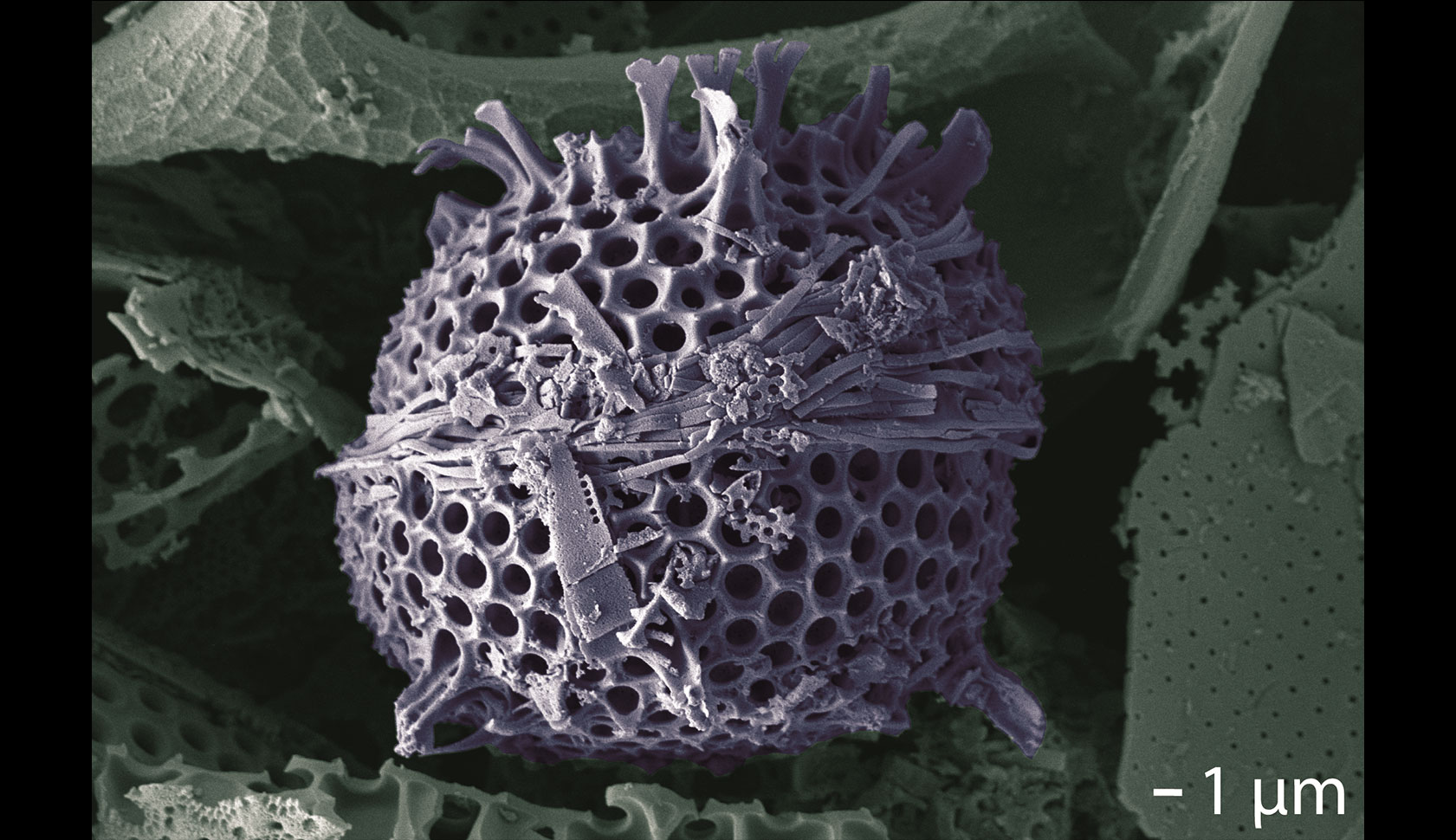
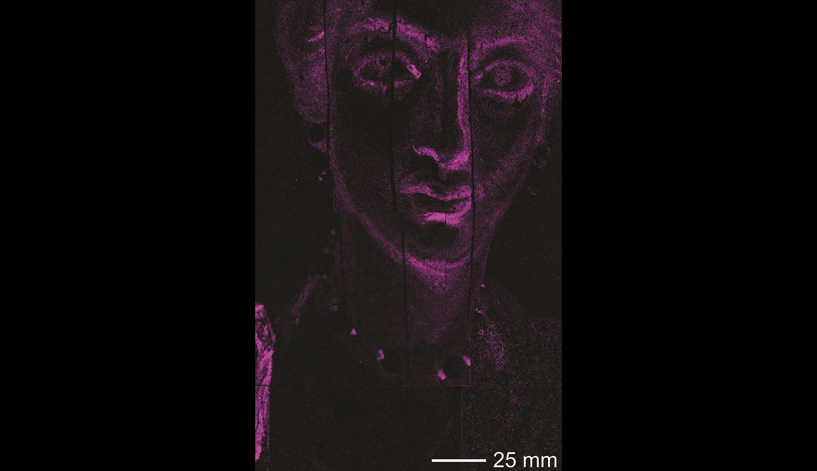
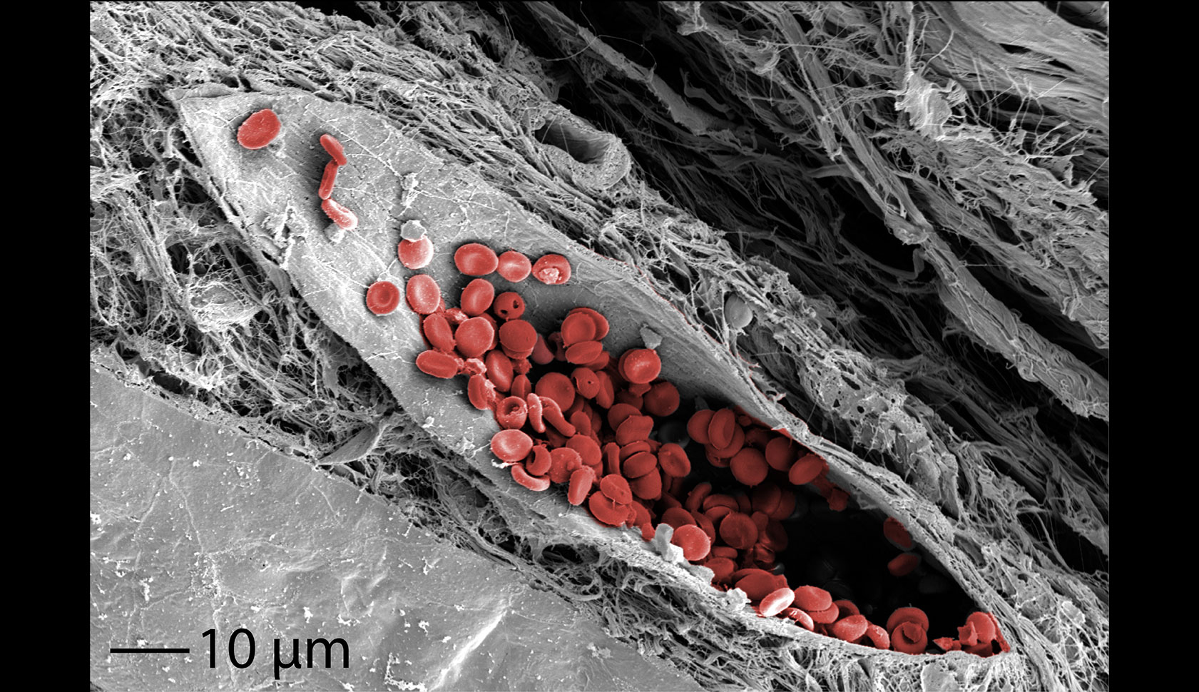
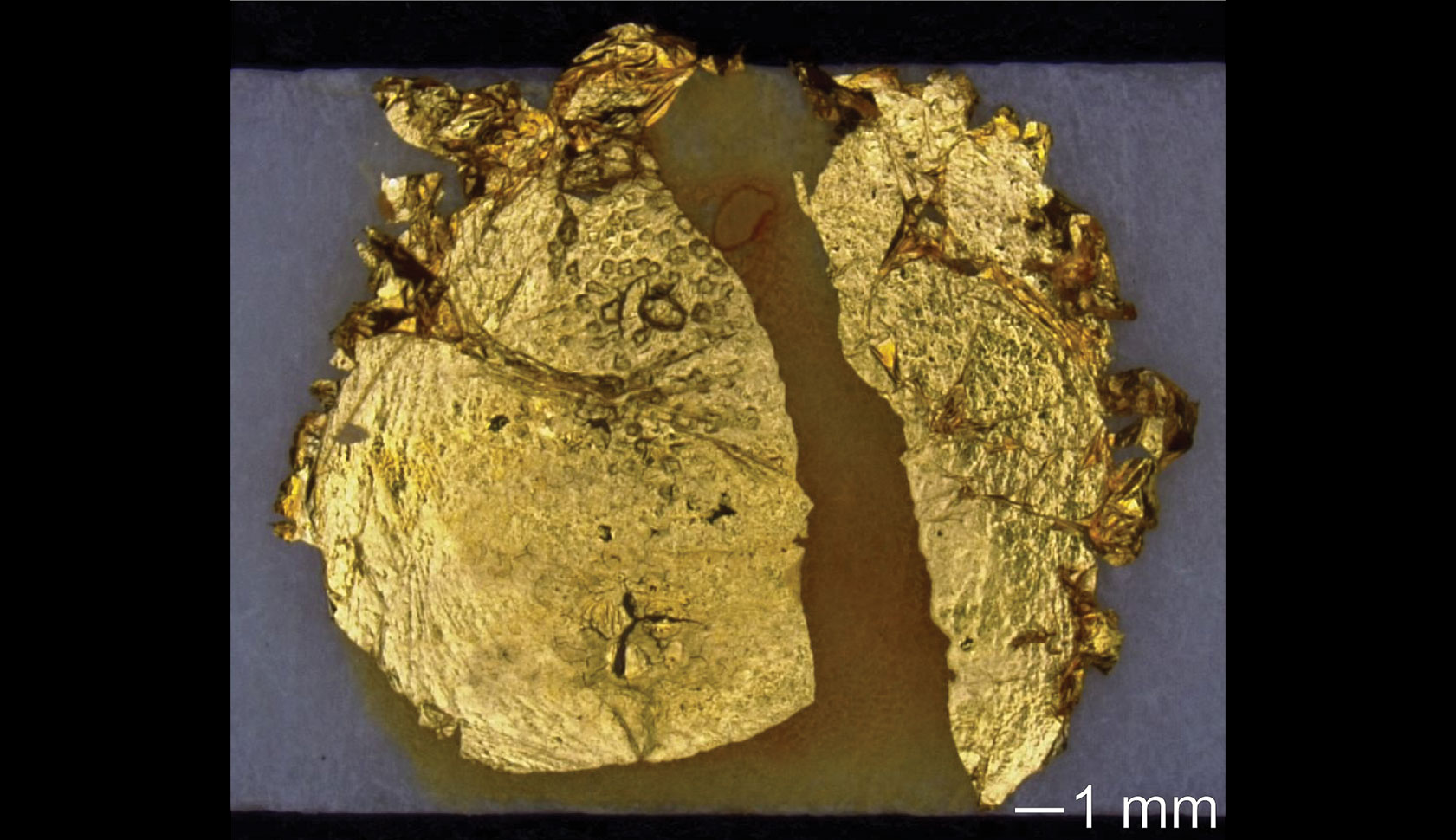
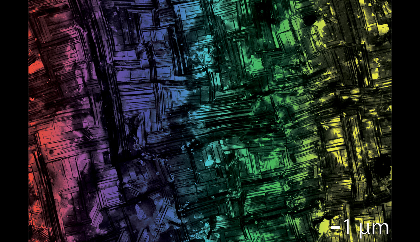
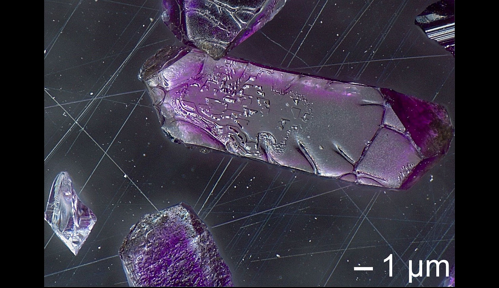
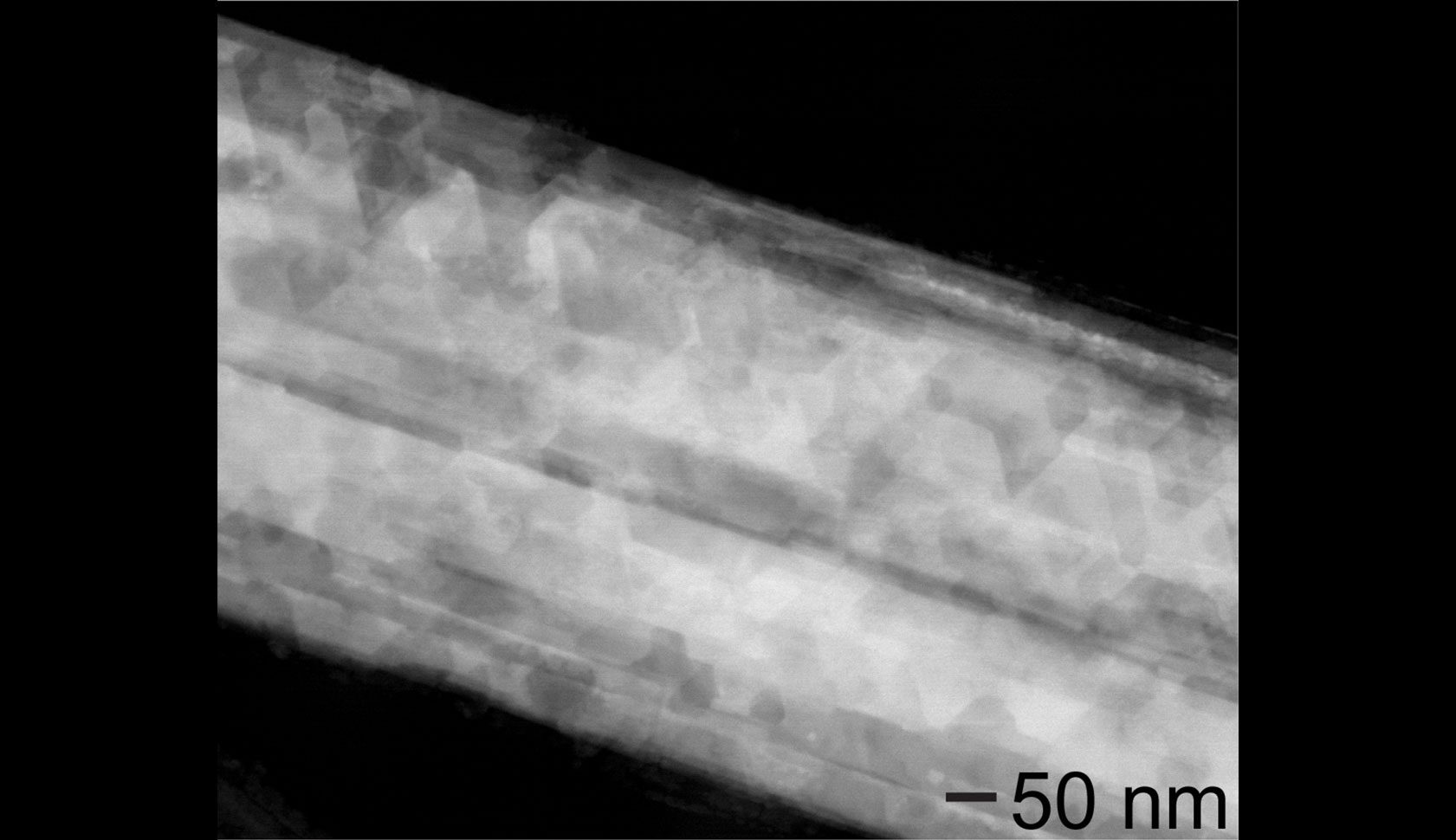
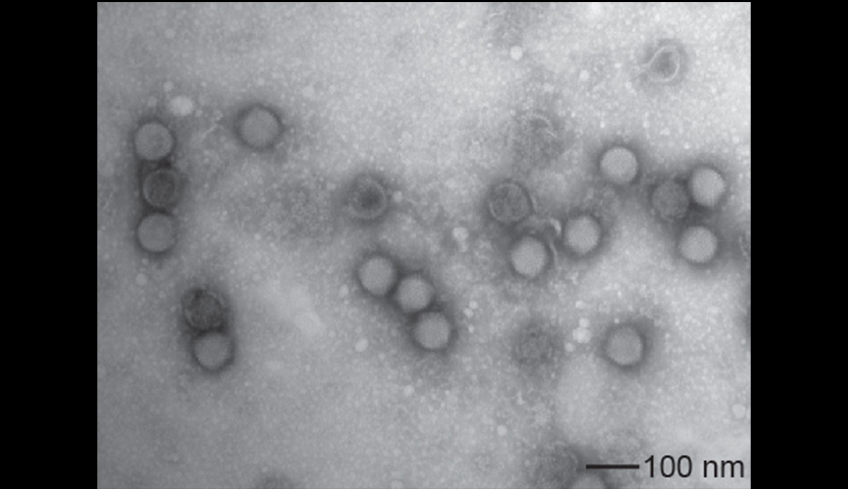
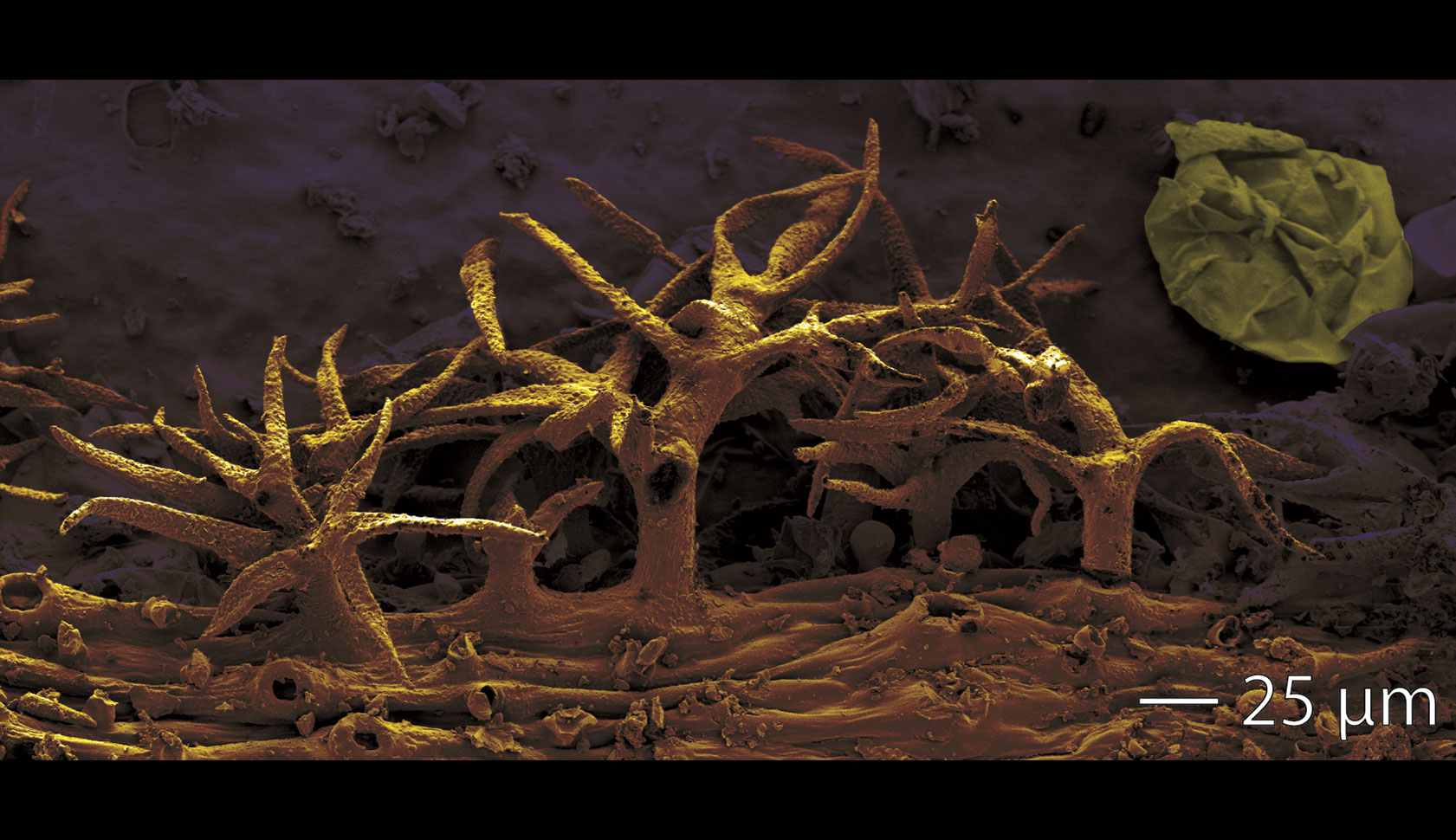
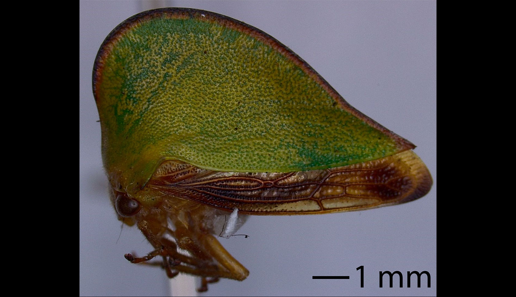
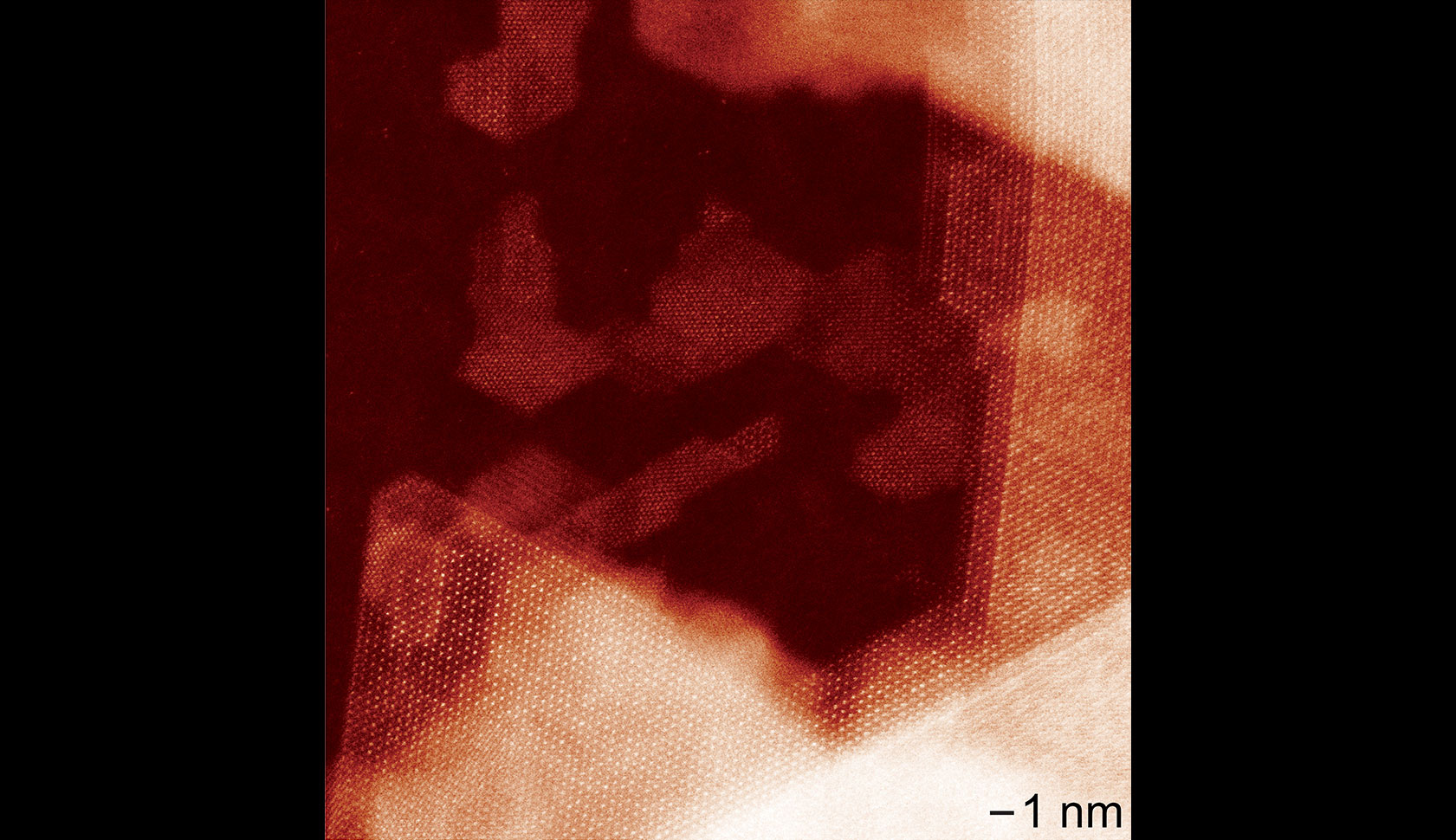
GEM has generated a variety of images for research, diagnostics, training, education and outreach purposes. The examples in this slideshow come from projects that range from analyzing gold particles on ancient art to examining red blood cells in an animal’s ear, to conducting experiments directly within the microscope at temperatures up to 1,000 degrees Celsius, and beyond.
For reference of scale in the images, 1 millimeter (mm) equals 1,000 mm (micrometers) and 1,000,000 nm (nanometers).


