
Scientific illustration is everywhere. You’ve seen it in posters at your doctor’s office, brochures from the veterinarian, the pages of furniture assembly manuals and covers of biology textbooks, just to name a few. Not just any artist can create these images—it takes an illustrator with years of specialized training, some of which may be from the University of Georgia.
UGA is home to a unique post-graduate certificate program in medical illustration and one of just 14 undergraduate majors in scientific illustration recognized by the Association of Medical Illustrators. As the point where fine arts and hard science intersect, scientific illustration is emblematic of the kind of multidisciplinary education at which UGA excels.

A unique post-grad experience

Offered through the College of Veterinary Medicine’s Educational Resources Center, UGA’s post-graduate certificate in comparative medical illustration provides students with classroom and experiential education in a variety of media. The program is helmed by Jim Moore, director of Educational Resources and a Distinguished Research Professor in large animal medicine.
While they take some graduate-level courses through the College of Veterinary Medicine, the students are focused on their assistantships in Educational Resources, where they work alongside medical illustration experts to put their training to the test. With this approach, their hard work amounts to more than a grade: Their illustrations help students at UGA and beyond.
“Our graduate assistants work with faculty members to create educational materials for use in their curricula, converting complex or difficult-to-envision concepts into visually appealing illustrations, animations or interactive 3D models,” said Moore. “It is our hope that these will help students develop strong mental models they can rely on when it comes time to apply these concepts later on.”
In addition to specific projects for professors at UGA, the Educational Resources team has created more than 60 free interactive eBooks covering a variety of topics that have been downloaded more than 140,000 times in more than 50 countries. Their expansive portfolio also includes several other kinds of media, including graphic design, 2D and 3D animation, photography and even game development.
Moore’s staff of illustrators, developers and photographers primarily work with faculty members within the College of Veterinary Medicine, giving the graduate assistants focused experience in illustrating the anatomy, physiology and treatment of animals large and small. But faculty across campus can request the team’s services, so they occasionally work on projects in other disciplines as well.

Pederson worked with undergraduate students in UGA’s Bachelor of Fine Arts in scientific illustration program to create 3D models—including this ant by Rachel Laird—that were displayed by the Russell Special Collections Libraries during an exhibit on invasive insects in Georgia. The B.F.A. program is housed in the Lamar Dodd School of Art, part of the Franklin College of Arts and Sciences. (Photo by Rachel Laird)
The program’s latest set of graduate assistants earned their certificates in May, following years of dedicated study in scientific and medical illustration. For Madison Christian and Linden Pederson, the combination of education and real-world application has been a valuable part of the program.
“The certificate is like a halfway step between graduate school and a real job,” said Christian. “It’s been good practice to talk to our clients, and there aren’t a lot of medical illustrators who have this certification, so it’s something that makes you stand out when applying for jobs.”
Christian’s education has involved more than just illustration classes and internship work. She and Pederson have observed surgeries at the College of Veterinary Medicine, and in their master’s program at Augusta University, they took anatomy classes alongside future doctors. This biomedical foundation is critical, because without accuracy and careful composition, medical illustration can’t serve its purpose of educating its viewers.
“The importance of each and every illustration being entirely accurate is something that’s really impressed upon us throughout our whole career as a scientific and medical illustrator,” said Pederson. “These are things that doctors will be using to explain anatomy to patients.”
And what about using photographs instead? Educational Resources’ medical illustrators often overlay their depictions of internal organs or surgical procedures on photographs taken by the unit’s medical photographer. The combination of photographs and medical illustrations works well to help patients, medical students and veterinary students more fully understand the material.
“If you were to use a picture from a surgery, you wouldn’t be able to see anything. With medical illustration, you have to be accurate but you also have to tell a story that people can understand,” said Pederson.
Learning how to tell this story and marry accuracy with effectiveness is the ultimate goal of medical illustration education, and a key part of the foundational training offered by scientific illustration bachelor’s programs.

Science in the art school
UGA’s Bachelor of Fine Arts in scientific illustration is housed in the Lamar Dodd School of Art in the Franklin College of Arts and Sciences. The program has produced decades of alumni who have gone on to pursue careers and graduate study in the field, including Christian, Pederson and its very own Chair of Scientific Illustration, Gene Wright.
Like all art majors, scientific illustration students learn about perspective, light, color and the other foundational components of creating any kind of art. But, while their peers in other specialties may experiment with impressionism or abstract art, scientific illustration students receive specialized training in depicting the world realistically.
“What we’re trying to do is give undergraduate students a fundamental understanding of what it means to be a science illustrator, and that includes the philosophy about how you’re supposed to treat this type of illustration,” said Wright. “It’s not about its beauty as an illustration; it’s meant to teach something. How do you lead a viewer through the drawing to teach them what you want them to learn?”
This lesson isn’t an easy one for Wright’s students. Before entering the major, many are more familiar with traditional art, where accuracy often matters less than color or emotional quality. As they work through projects—and endure several rounds of critiques—they begin to develop a thick skin and, more importantly, a scientific eye.


(Left to right) For one project, the B.F.A. scientific illustration students were asked to accurately measure and describe the scale, shape and form of found objects. Olivia Martin illustrated a pinecone, an atlas of a deer skull and a shell. (Illustration by Olivia Martin) This render and accompanying sketch, by B.F.A. student Noah Brown, reveal the inside of a pomegranate. For this project, said Wright, “the students were to create cubed sections out of fruit to learn how to define an educational section of information, as seen from two cut sides.” (Illustration by Noah Brown) Pederson created this 3D model of a fly. (Photo by Linden Pederson) Scientific illustration student Alex Kontos selected the American goldfinch for a project focused on illustrating an animal within its environment. (Illustration by Alex Kontos)
Each project begins with an objective, detailing the goal of the illustrations. With that in hand, the students begin to research their subjects, including setting up scenes and taking photographs that reflect the lighting or depth scenarios they hope to capture. This research evolves into quick, thumbnail sketches to help the students solidify their vision, and eventually final sketches.
At this point, the illustrations aren’t finished, but the students’ sketches will be scrutinized to determine how accurate they are and how well they meet the objective. After any adjustments are made, they move on to create the final illustrations. While students once used traditional artistic techniques like watercolor, these days they mostly use digital programs like Adobe Photoshop, Adobe Illustrator and Procreate.
With the illustrations complete, the students present their work for a final critique. After receiving feedback—and perhaps making a few more edits—the students are on to the next project. It’s an intensive process, but one that is necessary to achieve the level of accuracy scientific illustration requires, and to ensure that the students are getting a valuable experience.
It’s this detail-oriented education that leads the scientific illustration program’s alumni to success. Working with a mentor like Wright is an invaluable asset, even years after their graduation.
“Every time I’m working on a project, I hear Gene in my mind telling me, ‘That’s not right,’” said Christian. “I try to look at my projects through his eyes now—he really prepared me to produce the best work that I can.”

Pederson created this image for the cover of a supplementary issue of the Journal of the American Veterinary Medical Association. It depicts a caudal thoracic paravertebral nerve block. (Illustration by Linden Pederson)
Illustrating the future
Like the fields of science and medicine themselves, scientific illustration is ever-changing and evolving with the latest discoveries and technology—and UGA’s programs are keeping up.
Earlier this year, the Russell Special Collections Libraries hosted an exhibit on invasive insects in Georgia, featuring 3D models of the species created by Pederson and scientific illustration students. They used ZBrush, a digital sculpting program, to form each model, transforming a virtual ball of clay into a 3D figure. They then 3D-printed each one at a large scale and used traditional art techniques to transform the giant pieces of plastic into realistic insect models.
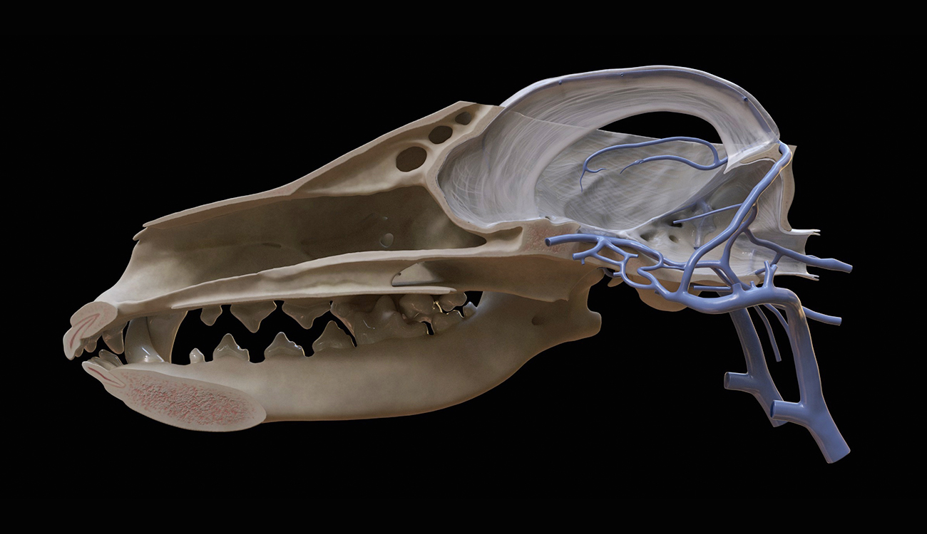
In medical illustration, the 3D modeling ZBrush offers is quickly becoming a standard approach. Through the graduate certificate program, students like Christian and Pederson have gained valuable experience with this cutting-edge technology. Among many 3D projects, Christian has modeled the inside of a horse’s hoof, and Pederson has visualized the veins of a dog’s brain.
It’s through projects like these that the graduate assistants hone their skills in visual storytelling and take their already well-developed talents to the next level. Certificate alumni enjoy success across the country, putting their training to work for hospitals, universities, biotech companies, journals and more. For many of them, a B.F.A. in scientific illustration from UGA was the first step in their educational journey, giving them the tools to stand out in their master’s programs and earn their way back to Athens for the certificate.
With both postgraduate and undergraduate programs in the field, UGA is on the forefront of innovation in scientific and medical illustration. Its students have learned to take both a scientific approach to art and an artistic approach to science, creating images that are accurate and tell a compelling story. They go on to provide all kinds of people—from young children to highly specialized physicians and researchers—with an accessible window into scientific subjects or processes.
Through their work, scientific and medical illustrators make science understandable for all.